
Ophthalmology
Eye clinic software
An eye exam writes the two eyes separately and reads them side by side. HekimBis keeps the exam in two panels, right (OD) and left (OS): visual acuity, refraction, tonometry, anterior and posterior segment findings. Measurements from devices such as the autorefractor, tonometer and OCT are carried into the exam record.
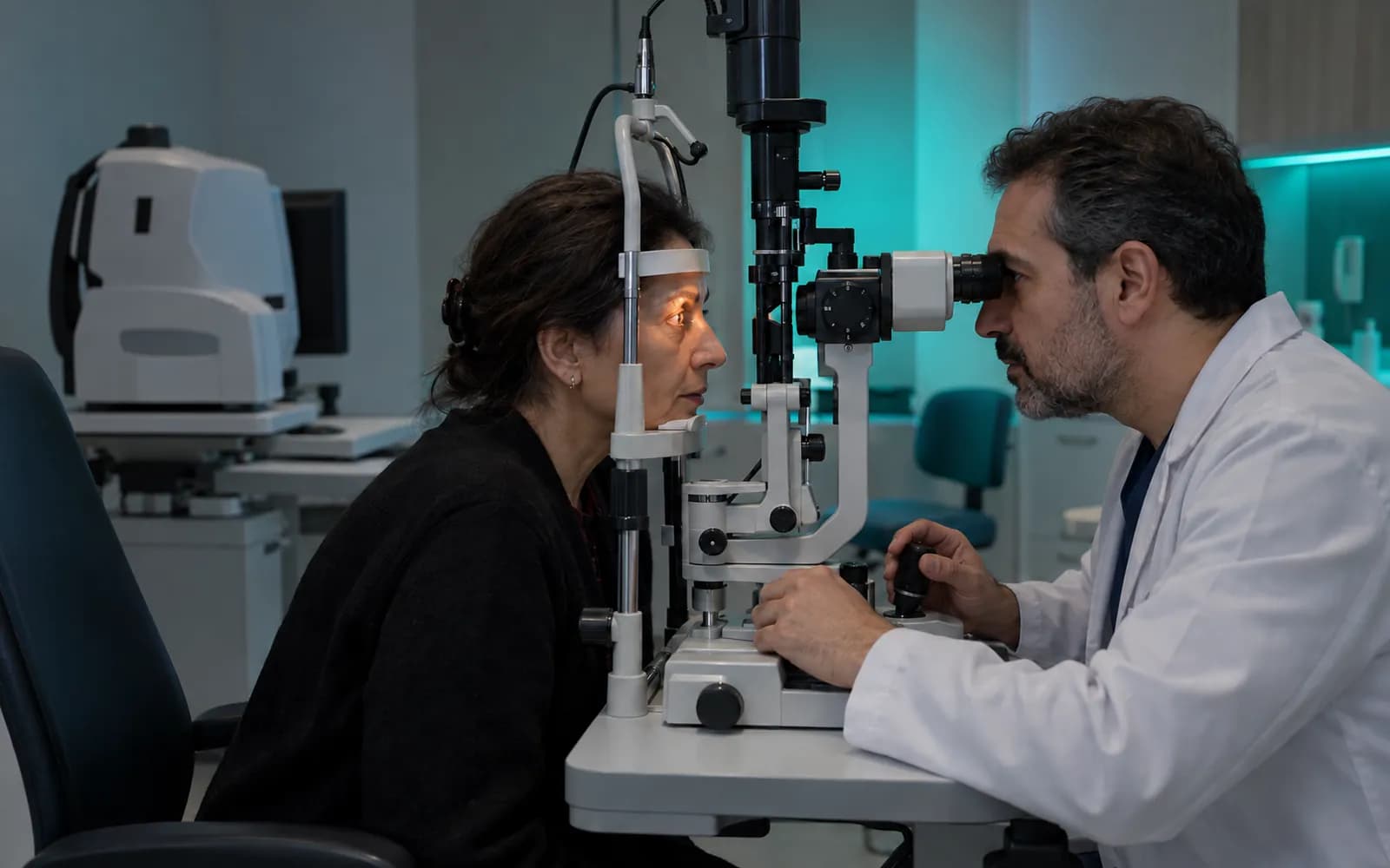
Measurements are checked, not typed
In an eye clinic the patient passes through several devices before the exam, and the real loss of time is getting the measurements onto your screen. The workspace matches the measurements to the patient and places them in the right field of the exam record; you check and interpret instead of typing.
When a patient at a routine check-up is measured with the autorefractor and tonometer, the values drop into the OD and OS panels of the exam record. You enter visual acuity, mark anterior and posterior segment findings and compare the OCT and fundus images with the earlier capture; a glasses prescription draft is prepared and a follow-up reminder is scheduled.
The right and left eye are written side by side on the same rows
On the two-panel screen, OD and OS line up on the same rows: visual acuity (distance and near, uncorrected and corrected), refraction values, tonometry and the anterior and posterior segment chart. Values that differ are highlighted and followed as a series between visits. On the chart you mark a finding and attach the OCT or fundus image.
| OD (right) | OS (left) | ||
|---|---|---|---|
| Distance vision | 0.9 | 0.9 | Manually entered |
| Near vision | 1.0 | 1.0 | Manually entered |
| Sphere | -1.00 | -1.50 | Measured by device |
| Cylinder | -0.50 | -0.50 | Measured by device |
| Axis | 175 | 10 | Measured by device |
| Intraocular pressure | 18 | 21 | Measured by device |
| Anterior segment | Normal | ! | Manually entered |
| Posterior segment | Normal | ! | Manually entered |
Intraocular pressure · Image linked
Differing values are highlighted; pressure of 21 and above is flagged.
Table view
| Value | OD (right) | OS (left) |
|---|---|---|
| Distance vision | 0.9 | 0.9 |
| Near vision | 1.0 | 1.0 |
| Sphere | -1.00 | -1.50 |
| Cylinder | -0.50 | -0.50 |
| Axis | 175 | 10 |
| Intraocular pressure | 18 | 21 |
| Anterior segment | Normal | ! |
| Posterior segment | Normal | ! |
1
Names, values and records on screen are examples prepared for illustration.
Four needs of an eye clinic
- Visual acuity and refraction
- Distance and near vision, corrected and uncorrected values and refraction values are followed as a series.
- Tonometry and follow-up
- Intraocular pressure measurements are followed as a line across visits; the check interval is tied to the plan.
- Anterior and posterior segment
- Findings are recorded with a schematic drawing and defined terms; the right and left distinction is kept.
- Imaging and field tests
- OCT, fundus and perimetry results attach to the patient card as images and reports.
OCT and fundus images compared over time
The earlier and current OCT or fundus image of the same eye opens side by side; you mark the region and add your note. Images match the patient and the eye (OD or OS), and access is limited by role and care relationship.
- Earlier and current image side by side
- Region mark and note
- The image matches the patient and eye (OD/OS)
- Access in the audit log
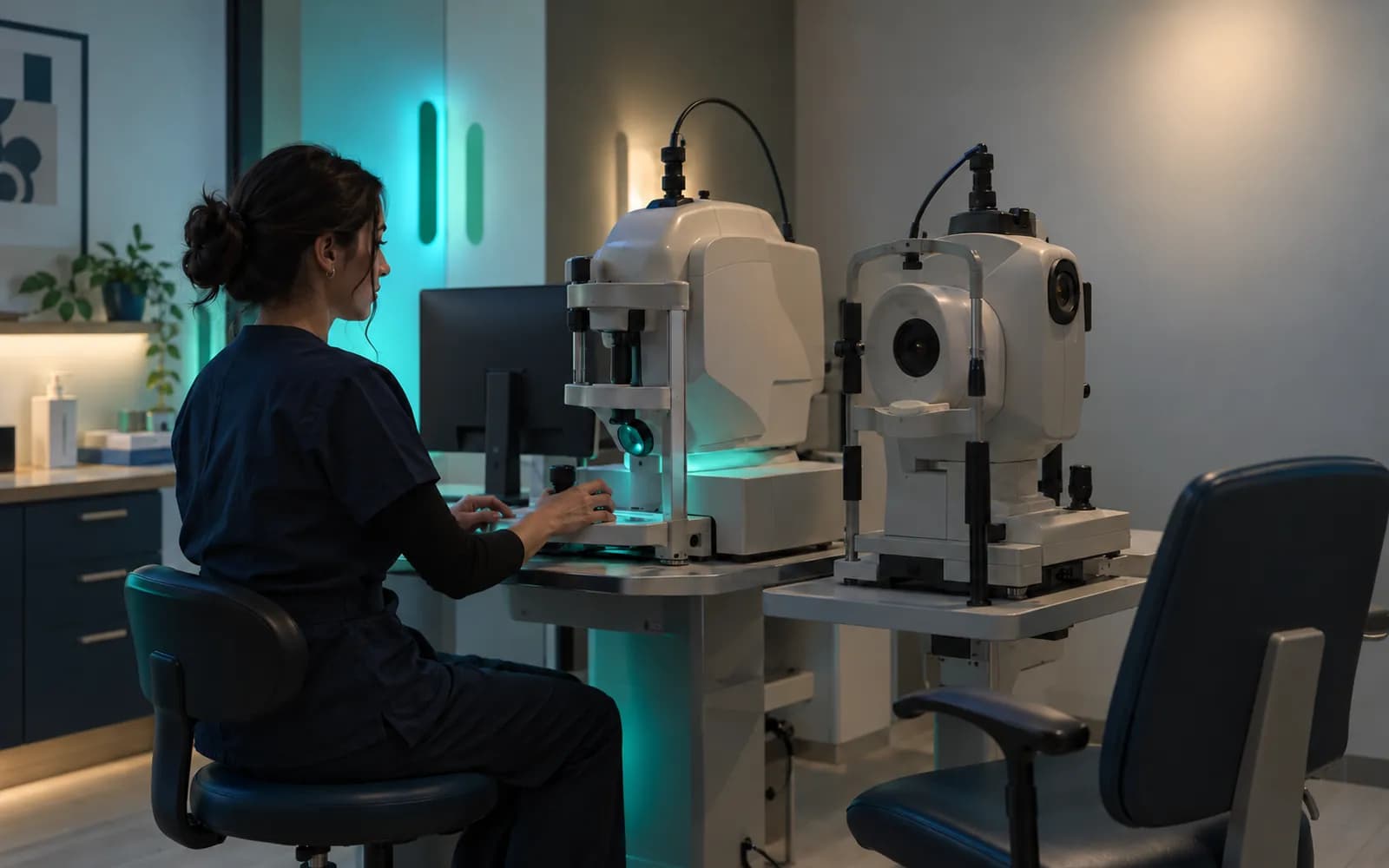
From the device to the exam, without retyping
Values from measurement devices are matched to the patient's identity and settle into the relevant OD or OS field of the exam record; a result that does not match does not attach on its own and falls to human review. If the device connection drops, a local queue keeps the values and re-matches them when the connection returns; the same measurement does not create a duplicate record.
- Matching by patient identity
- A mismatched result goes to human review
- The local queue keeps values during an outage, with no duplicate records
- A value entered by hand is marked
Record safeguards in the eye exam
- Matching to the patientEvery measurement is matched to the patient; a result that does not match is not attached automatically.
- Source markingValues from a device and values entered by hand are marked separately.
- Right and left eyeRight (OD) and left (OS) eye values are kept in separate fields.
- Approved prescription draftA glasses prescription draft does not become final until you approve it.
- An outage-tolerant queueIf the connection drops, a local queue keeps the values and matches them again when the connection returns.
Device flow in an eye clinic
An eye clinic has the most devices of any specialty; measurements attach to the exam record and images to the patient card.
- Autorefractor and keratometer
- Refraction measurements drop into the exam record.
- Tonometer
- The intraocular pressure value enters the record.
- OCT
- The image and report link stay on the patient card.
- Fundus camera
- The image is added to the archive.
- Perimetry
- The visual field report attaches to the file.
Common questions about eye clinic software
Is there a separate profile for ophthalmology?
Yes. Ophthalmology is one of the defined clinical profiles: OD and OS, visual acuity, refraction, tonometry, anterior and posterior segment and follow-up. It is versioned and passes two separate clinical sign-offs before release.
Are my devices supported?
Device connections are verified per manufacturer, exact model, protocol and version, and the verified ones are listed. An image or value file from a device that is not on the list can be imported with a note that it is not integrated.
How is a glasses prescription prepared?
A glasses prescription draft is produced from the refraction values; you edit and approve it.
How are OCT images used?
The earlier and current image open side by side, and the HekimBis viewer is for clinical assessment and comparison.
How does it work for child patients?
The parent and guardian record and parent communication are defined on the patient card; messages carry no health data.
Are there follow-up intervals and reminders?
Tonometry and other check intervals are tied to the plan; patients who are due fall onto a list, and a patient who does not come generates a task.
How are surgery and consent records kept?
Procedure-specific clinical consent is a record separate from disclosure and explicit consent; a signed consent does not change, and a correction is an addendum whose history is kept.
What about areas such as oculoplasty and aesthetics?
This profile is for the eye exam and follow-up. For aesthetic procedures, the Medical Aesthetics and Plastic Surgery profiles are defined separately and can be used together.
Keep reading
Related pages
Features
- Device connectivitySecure, outage-tolerant data from clinic devices through the Edge Connector.
- Imaging and PACSDICOM workflow from order to report: worklist, matching, and a reference viewer.
- Multi-branchBranch-level permissions, prices, stock, and cash with central call handling.
- Front desk and check-inArrival logging, QR or kiosk check-in, waiting room status, and delay estimates.
Specialties
Software guides