
Dermatology
Dermatology clinic software
In skin disease, the decision often depends on how a lesion changes over time. HekimBis places the lesion on a body map, records its morphology in defined fields and links photos and dermoscopy images to the same lesion, so every visit puts the new image beside the previous one.
A lesion record takes two touches
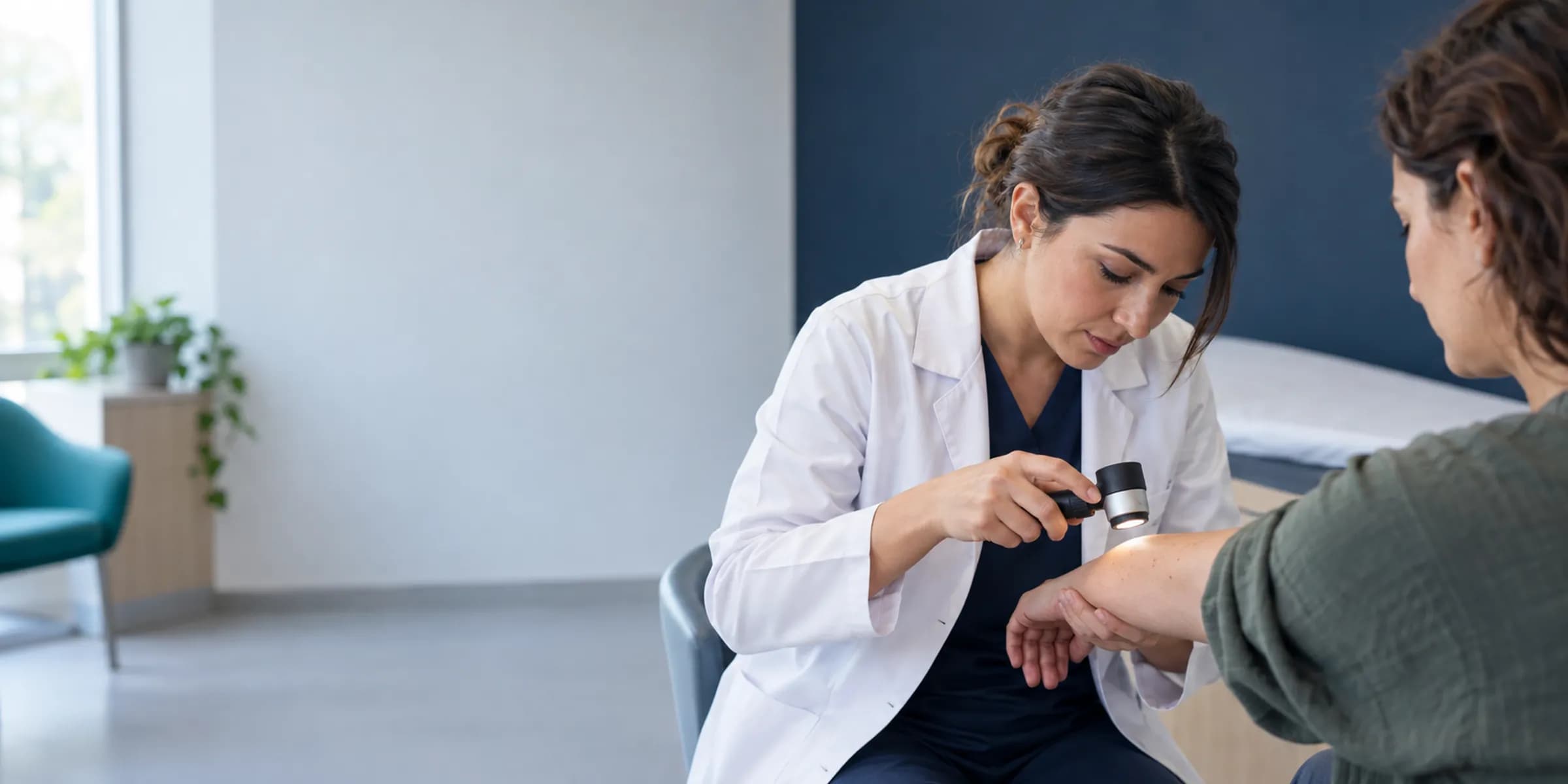
In the exam room you hold a dermatoscope and have no time to wrestle with a screen. So the lesion record stays short: pick the site on the map, enter morphology and size, and the image attaches to the lesion when you capture it. At the next visit the same lesion of the same patient is waiting, marked on the map.
For a patient with several moles, you mark each lesion on the map, write its size and attach the dermoscopy image. At the check-up six months later the map shows the same sites, and you select lesions and compare them with earlier images. For a lesion that has changed, a procedure plan and a pathology order are opened, and the result attaches to the lesion record when it arrives.
On the body map, every lesion is a marker
On the front and back body silhouette, each lesion appears as a marker. Tap one to open its morphology fields (color, border, surface, size), measurement, date and the linked photos and dermoscopy images. The time slider shows the map as it stood at past visits and highlights the lesion that changed; an outside report or pathology result attaches to the lesion.
1 · Under follow-up · Front
- Colour
- brown
- Border
- regular
- Surface
- flat
- Size
- 4 mm
Pathology linked · Photo available
Table view
| # | Status | Region | Colour | Border | Surface | Size |
|---|---|---|---|---|---|---|
| 1 | Under follow-up | Front | brown | regular | flat | 4 mm |
| 2 | Changed | Front | dark brown | irregular | raised | 6 mm |
| 3 | Under follow-up | Back | pink | distinct | scaly | 9 mm |
| 4 | Procedure done | Back | red | distinct | crusted | 5 mm |
Names, values and records on screen are examples prepared for illustration.
Three layers of a dermatology visit
- Morphology and lesion record
- Lesion, site and size are entered in defined fields, producing comparable data instead of free text.
- Clinical photo protocol
- The angle and order of photos are defined, and the dermoscopy image attaches to the lesion.
- Treatment plan and serial follow-up
- Medication, procedure and check interval are kept as a plan; a patient who does not come generates a task.
The same lesion, a different visit
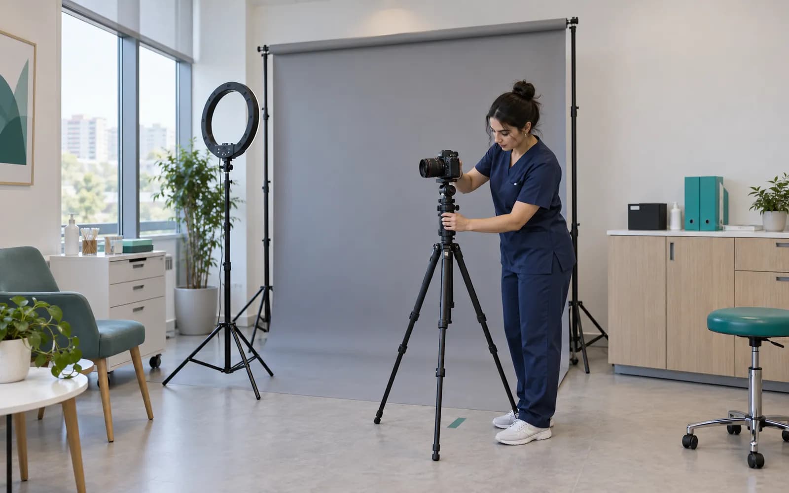
Serial photos taken at a standard angle and light are shown side by side or overlaid. Photos serve the clinical record; a separate photo consent is obtained for sharing or promotional use, and without a consent record the image is not offered for sharing. Interpreting the comparison belongs to the physician.
- The previous visit and the current image are compared with a scale
- Photo consent is visible in the record
- An outside report or pathology result attaches to the lesion
For patients from abroad
Patient surfaces are available in Turkish, English, Arabic, Russian and German, and the team screen runs in Turkish and English. Form and consent translations are approved by people. Remote pre-assessment and quotes in foreign currency are part of the health tourism capability.
- Türkçe
- English
- العربية
- Русский
- Deutsch

Dermatology device surface
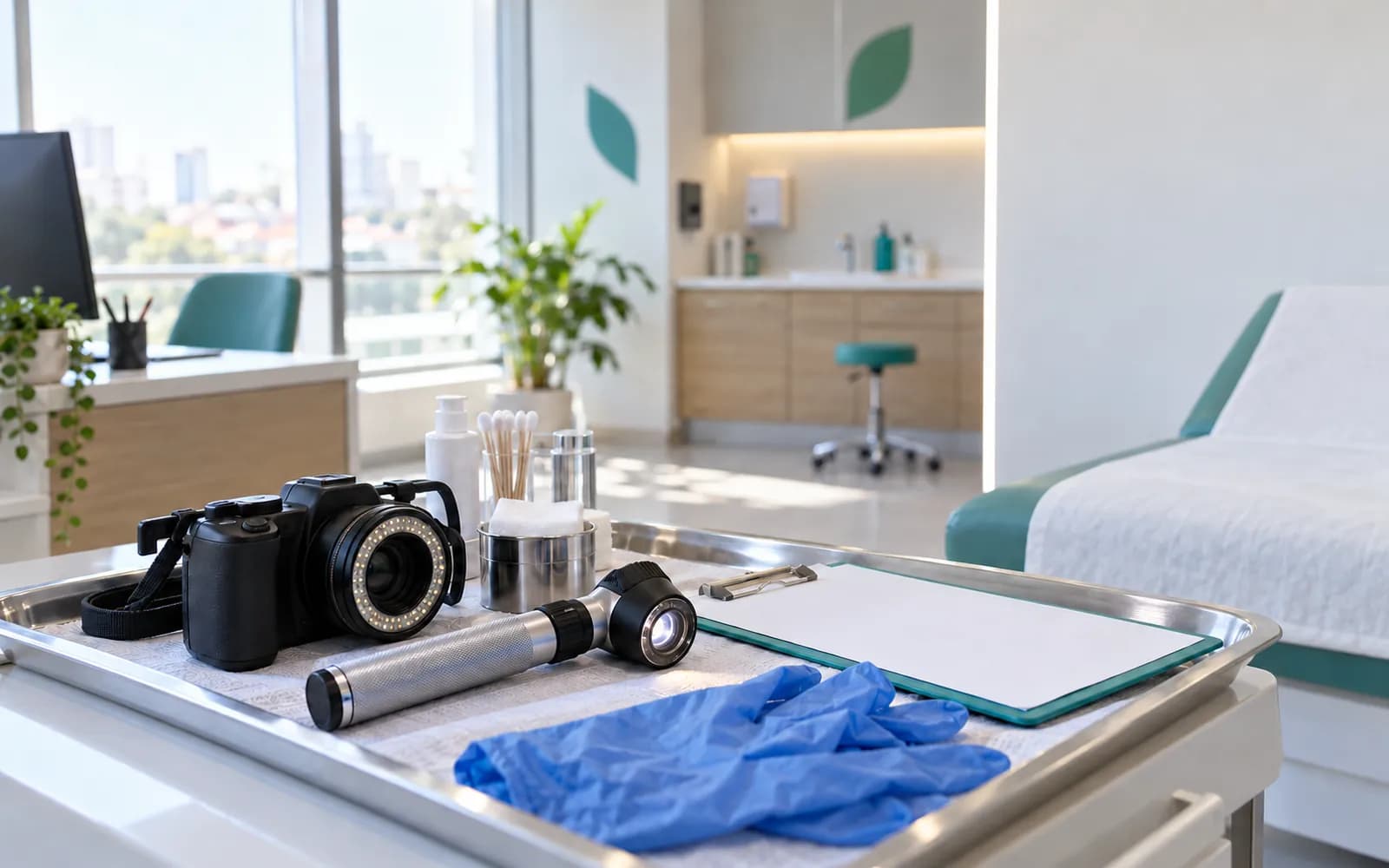
Dermoscopy and standard photography are the main image sources in dermatology, and the images attach to the lesion and the visit.
- Dermatoscopes
- The image attaches to the lesion.
- Standard clinical photography
- Added as an ordered series following the protocol.
- Skin ultrasound
- The image and measurement file are added to the record.
- Procedure device outputs
- The session summary enters the file.
Common questions about dermatology software
Is there a separate profile for dermatology?
Yes. Dermatology is one of the defined clinical profiles: lesion and body map, morphology, serial follow-up, clinical photo protocol and treatment plan. It is versioned and passes two separate clinical sign-offs before release.
Where is AI used?
AI only produces note drafts, and a draft does not become final until you approve it. Assessing the lesion and the diagnosis belong to the physician; image and measurement comparison is a recording tool that supports that decision.
Are photos kept safely?
Photos are part of the patient file; access is limited by role, branch and care relationship, and every access is written to the audit log. Sharing needs a separate consent.
Is my dermatoscope supported?
Device connections are verified per manufacturer, exact model, protocol and version, and the verified ones are listed. An image file from a device that is not on the list can be imported with a note that it is not integrated.
Are aesthetic procedures part of this profile?
Medical aesthetics and plastic surgery are separately defined profiles. A clinic can use dermatology together with them.
Who sets the serial follow-up intervals?
Check intervals come from the protocol in the clinical definition, and your clinic can add its own tasks and reminders. The safety and clinical rules in the central definition stay in place.
How does a pathology result attach to a lesion record?
A pathology order is opened for the specimen taken during the procedure; when the result arrives, the order, specimen and lesion match under the same identity. A result that does not match falls to human review.
Keep reading
Related pages
Features
Specialties
Solutions and process
Software guides