
Imaging, DICOM and PACS workflow
An image is more than a file; it belongs to an order, a patient, a device and a report. HekimBis follows every step from the order until the report reaches the patient, with its status, time and owner. An image coming from a device is matched to the right patient and the right order, and if it cannot be matched it is quarantined and an authorized user decides.
The device takes the patient from the worklist
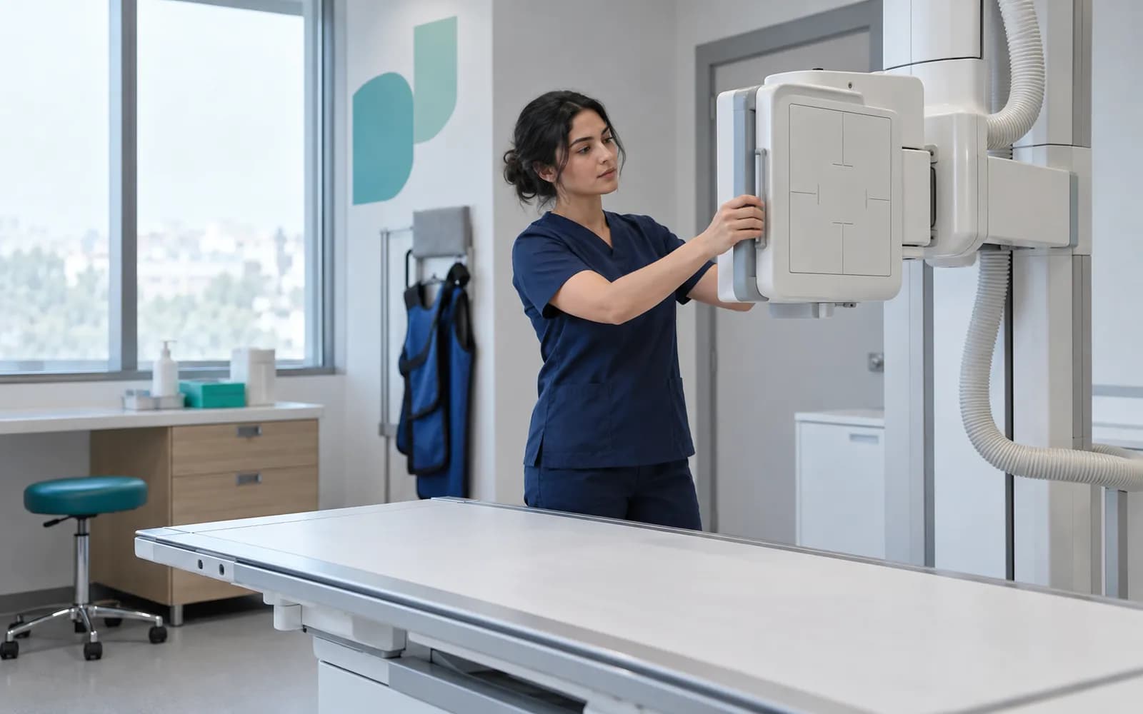
When the physician opens an imaging order, it reaches the device's worklist with an accession number and the patient's identity. The technologist picks the patient from the list, so there is less need to retype patient details on the device.
When the exposure is finished, the image is transferred to the archive securely and matched to the order and the patient. An image that does not match, arrives twice or arrives late is quarantined, and because a name alone is never grounds for matching, two patients with the same name are not mixed up.
In outpatient organizations and small and mid-sized diagnostic centers, the radiology workflow runs through these steps from order to report, including preparation checks, scheduling, the exposure, technical quality control, repeat exposures and rejection reasons.
An imaging order from scheduling to the portal
The physician opens the order, the device takes the patient from its worklist, the exposure ends, and the image is transferred to the archive and matched to the order. The report is signed, a critical finding is communicated to the physician and the approved result is released in the patient portal. An image that does not match is quarantined, and repeat exposures and addenda are followed as separate branches.
Timeline and portal
The report, key image and study are added to the patient timeline and, with an authorized user's approval, published in the patient portal.
- The process is complete here.
- Study 0007
- Series 2
Published
The names, times and records on the screen are examples.
All steps
- 01 Imaging order (Clinic) The physician opens the order: clinical reason, body site, exam type and priority. Contrast and allergy checks are made if needed.
- 02 Accession and worklist (Planning) The order receives an accession number, is tied to an appointment and room, and appears on the device worklist.
- 03 Arrived and started (Modality) The patient has arrived, the device selects the patient from the list and the exam begins.
- 04 Exam completed (Modality) The device reports the exam is finished; a technical quality check is made. If inadequate, a repeat exposure.
- 05 Transfer and technical check (Archive) The image is transferred to the archive; count, integrity and format are checked.
- 06 Patient and order matching (Archive) The image header is compared with the patient and order identity. If it matches it enters the reading queue.
- 07 Reading queue (Reporting) The study waits in the reading queue by specialist assignment and priority.
- 08 Draft and final report (Reporting) The report is written from a template, kept as a draft, signed and made final. The system on which the primary interpretation was made is recorded.
- 09 Critical-finding notification (Reporting) If there is a critical finding, the ordering physician is notified; an acknowledgement is awaited and escalated if absent.
- 10 Timeline and portal (Portal) The report, key image and study are added to the patient timeline and, with an authorized user's approval, published in the patient portal.
Alternative path from 04 Exam completed; it returns to the main flow at 03 Arrived and started.
- 04a Repeat exposure (Modality) If quality is inadequate or there was movement, the exam is repeated; the rejection reason is recorded and the exam starts again.
Alternative path from 06 Patient and order matching; it returns to the main flow at 07 Reading queue.
- 06a Quarantine: header did not match (Archive) The patient or order identity does not match, or the image is a duplicate or late. The image goes to quarantine; no automatic matching is made.
- 06b Human review (Archive) An authorized user matches the image to the right patient and order or rejects it; the decision is recorded and the image enters the reading queue.
Alternative path from 08 Draft and final report; it returns to the main flow at 10 Timeline and portal.
- 08a Addendum / correction (Reporting) If information must be added to a final report, an addendum is opened; if there is an error, a correction. The previous version is kept and the current version reaches the patient timeline.
10 Timeline and portal
Six ways the device and the archive talk to each other
HekimBis talks to devices through standard DICOM services. They define how a device connects to the archive, how it receives the patient and how it hands over the image.
- C-ECHO, connection test
- Confirms that the device and the archive can reach each other.
- C-STORE, image transfer
- Studies, series and images are accepted into the archive.
- Modality Worklist
- The device takes the patient from the system, so retyping patient details is needed less often.
- MPPS, exposure status
- The device reports when the exposure has started and finished.
- Storage Commitment
- On devices that support it, the archive confirms to the device that the image is safely stored.
- DICOMweb, web-based access
- Query, retrieve and store through QIDO-RS, WADO-RS and STOW-RS.
Where needed, querying and retrieval also run through C-FIND and C-MOVE. Transfers are tested for SOP class, transfer syntax, character set and vendor private tag compatibility.
DICOM and PACSAlongside an existing PACS, or with a managed archive
Connection to an existing PACS or RIS
If your organization already runs an enterprise PACS or RIS, HekimBis works alongside it. The imaging order, status, study information and report are managed in HekimBis, and an authorized, audited handover opens the viewer. Pixel data is not duplicated unnecessarily, and the order, status, study and report are reconciled between the two systems.

The HekimBis managed archive
For outpatient organizations and small and mid-sized diagnostic centers, HekimBis offers its own managed archive. Image data is stored separately from the clinical record, encrypted and isolated by organization and branch. Retention and lifecycle are defined, and integrity is verified with checksums. Thumbnails, an authorized web viewer and study comparison are included, measurements and annotations are tracked with the image and the user, and sharing and export are written to the audit trail. Archive capacity is added as extra capacity; see the pricing page for details.
A report moves from draft to final, addendum and correction
A report is written with a template specific to the specialty and the type of exposure. It has draft, signed and final states.
If information is added later an addendum is opened, and a mistake is handled with a correction or a withdrawal while the earlier version is kept. A study can be compared with a previous study and report, and the report, key image and study appear together on the patient timeline.
When a critical finding is detected, the physician who wrote the report communicates it to the ordering physician. Who received the notice and when, and the acknowledgement, are on record, and if it does not arrive the process is escalated.
The HekimBis viewer is labeled as a clinical and reference viewer. The primary interpretation is made at the radiologist's approved diagnostic imaging workstation, HekimBis provides an authorized, audited handover to it, and the report signing step records which system the interpretation was made in.

Frequently asked questions
Can I connect my X-ray device to HekimBis?
A device connection is verified for a specific manufacturer, exact model, protocol and software version, and only verified combinations are listed as supported. If an image file can be taken from a device that is not on the list, it is labeled as a manual import, not integrated. The current list is on the device connectivity and integrations pages.
Do I need to replace my existing PACS?
Your existing enterprise PACS or RIS stays, and HekimBis connects to it and reconciles the order, status, study and report. The managed archive is a separate working mode for outpatient organizations and small and mid-sized diagnostic centers.
How is the risk of attaching an image to the wrong patient prevented?
The information in the image header is compared with the patient and order identity, and an image is never linked automatically on name alone. An image that does not match, arrives twice or arrives late is quarantined and an authorized user decides. Cases such as two patients with the same name or a duplicate accession number are caught this way.
How does a critical finding reach the physician?
The physician who wrote the report communicates the critical finding, the notice goes to the target person with its channel and time on record, and an acknowledgement is awaited. If none arrives the process is escalated, and all steps appear on the patient timeline.
Does a result reach the patient portal automatically?
A result appears in the portal when an authorized user releases it. Even when a report reaches its final state, release is a separate, explicit action, which prevents early publication.
Is pathology covered too?
For small and mid-sized diagnostic centers the pathology order, specimen chain, report and critical finding flow are covered, and an authorized, audited handover opens an existing approved digital pathology viewer.
Are images lost if the connection drops?
The Edge Connector inside the clinic network keeps images and messages in an encrypted local queue during an outage and forwards them without repeats once the connection returns. See the device connectivity page for details.
Which steps does the radiology workflow go through?
The order, contrast and allergy check, scheduling and check-in, room and technologist assignment, the exposure, technical quality control, the reading queue, the report and critical finding notification are followed in turn. Repeat exposures and rejection reasons are also on record.
What does the web viewer offer?
The authorized web viewer offers thumbnails, study and series comparison, measurement and annotation. Measurements and annotations are tracked with the image and the user, and sharing and export are written to the audit trail.
Keep reading
Related pages
Features
Specialties
- RadiologyModality worklist, acquisition, matching, signed reports, and critical findings.
- Orthopedics and traumatologyRegion and implant cards, pre- and post-op follow-up, and linked imaging.
- DentalTooth and surface odontogram, treatment plans, prosthetics, and outside lab work.
- Obstetrics and gynecologyPregnancy timeline, ultrasound records, screening tests, and visit schedules.
Solutions and process
Software guides
Integrations